Arthritis

Osteoarthritis – not just a disease of older horses.

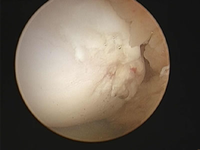
Osteoarthritis is one of the most common causes of lameness, poor performance and premature retirement in racehorses. It should not be considered as a problem just in “older” horses, as early stages of osteoarthritis can develop in horses as young as two years old.  Arthritis does not have to be debilitating for your horse.  Treatments are effective and are readily available including many new ways of managing this disease.

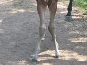
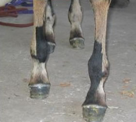
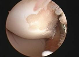
Trainers commonly report their horses to be “jarred up” particularly after a race or gallop on a hard track. These horses may not be lame, but are described as “a bit scratchy” or reluctant to “stretch out”. This may be perceived as “normal” for a particular horse but in fact, it may be an early warning sign of joint pain. Some horses may not show any signs of soreness at all, but develop swelling, filling, heat or pain around the affected joint. These are all common signs of joint inflammation. Joint inflammation can arise simply from repeated stress incurred during everyday training. Healthy joints are protected by cartilage, which acts as a shock-absorbing cushion between the bones, and a thick sticky synovial joint fluid, which acts as a lubricant. Joint inflammation causes the release of destructive enzymes into the joint, which if left untreated or allowed to recur repeatedly, have an irreversible destructive effect on the joint cartilage and the quality of the synovial fluid.

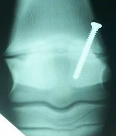
It is impossible to prevent joint stress however the aim is to normalise the inflamed joint as quickly as possible before permanent damage occurs. As osteoarthritis is not curable, the key to successful management and prevention of ongoing damage is early detection and prompt treatment. Routine management procedures such as icing and bandaging, anti-inflammatory drugs (phenylbutazone), anti-arthritic drugs (Cartrophen, Pentosan, Hyaluronic Acid etc.) and cortisone joint injections are effective treatments for managing osteoarthritis. Recently a new technique known as IRAP has been developed which involves collecting and using the horses’s own natural antibodies as a joint injection. If you have any further questions, please call the clinic and we will be happy to discuss this with you.

Angular Limb Deformities in Foals

carpal-valgus

Angular limb deformities (ALD) occur in foals and if left untreated may result in lameness and poor performance as adults. These deformities can be present at birth or develop as the foal ages. The most common sites for ALD’s are at the level of the growth plate such as the distal radius (carpus/knee) and distal cannon bone (fetlock).

The deformities or deviations are described as either valgus or varus and we most commonly see carpal valgus (knock-knees) and fetlock varus (toe-in). The treatment of ALD’s depends on the growth potential that is left at the affected growth plate. As foals age, the growth plate closes, with the lower limb’s growth plate closing earlier than the upper limb. For this reason, surgical treatment of ALD’s is often recommended when the foal is as young as 4 weeks of age. Surgical treatment is undertaken when the deformity is worsening or where it is not responding to conservative treatment.

Fetlock Varus

Often, stall or small yard confinement alone is sufficient to correct mild deformities. Confinement is usually carried out in combination with corrective trimming of the affected foot by your farrier. Your farrier and veterinarian will often evaluate your foal together to determine if the foal is improving with conservative therapy alone. Sometimes, your veterinarian will recommend surgical intervention in order to obtain the straightest limbs possible to prevent future complications such as lameness. The optimum time to perform surgery is during maximal growth at the level of the growth plate. Remember, once the growth plate closes, surgery is unrewarding and will not benefit the horse.

Periosteal stripping is a surgical technique that is performed frequently by surgeons around the world to correct ALD’s in foals. This technique involves making a keyhole incision through the skin and then incising the periosteum (sheath around every bone) to ‘release’ the tighter side (concave side) of the limb immediately above the affected growth plate. This is often performed on fetlock deformities at the age of 4 weeks and on carpal deformities (knees) at around 6 weeks of age because of the maximal growth potential at these times. This procedure is frequently sufficient to correct most mild deformities and results are generally rewarding.

physeal-screw

Occasionally, for more severe deformities or where periosteal stripping may not have achieved sufficient limb straightening, we may recommend that the foal undergo transphyseal bridging. This procedure is performed on the convex side of the affected growth plate where growth is faster than the other side of the limb. We insert a screw across the growth plate to stop growth on the convex side to let the slower side ‘catch up’. This is a more invasive technique and a second surgery is needed to remove the screw when the limb is straight to prevent over-correction. Transphyseal bridging is usually recommended for fetlocks when the foal is 3 months old. For knees, we tend to get a reasonable response even up to 12 months of age, however, if the deformity is severe, we will often recommend surgical intervention in the first few months of life. This surgical technique is highly effective for achieving limb straightening and is a routine procedure for many affected foals in breeding areas all over the world.

Yearlings at the sales are critiqued by many veterinarians and potential buyers. ‘Bent legs’ are a common reason for yearlings not achieving their optimum selling price because of the chance for future complications. In foals with ALD’s, early evaluation and intervention by your veterinarian and farrier will optimize your potential for presenting a horse with straight limbs and achieving the best selling price.

Arthroscopy – What do we see?

arthroscopy

Arthroscopic surgery makes up around 40% of all surgical procedures undertaken at GCEC. Similar to human ‘keyhole’ surgery, this technique allows us to perform a minimally invasive approach to evaluate joint surfaces and treat abnormalities such as osteochondral fragmentation (bone chips).

There has been much research into the cause of bone chips within horse’s joints. What we know is that the majority are caused by repetitive injury of the cartilage rather than being due to one acute episode. Cartilage is a shock-absorbing cushion of tissue that overlies bone within a joint and when it becomes damaged over time, it hardens and the bone beneath it becomes brittle leading to an increased chance of it breaking. Thus, when a surgeon is able to look at the cartilage during arthroscopic examination, they are able to provide a prognosis for future athletic activity based on the degree of damage to the articular surfaces.

athroscopy

What we are also learning is that we can manipulate the health of cartilage through chondroprotective agents given by injection, orally or both. These agents serve to enhance the quality of repair tissue that forms where cartilage defects are debrided and also to reduce inflammation which causes continued cartilage degradation. We often see trends in racing circles between regions of Thoroughbred racing and know that many stables in Flemington and Randwick as well as here on the Gold Coast are administering these products to promote soundness and maximize the number of starts for each horse.

We recommend that horses undergo a course of treatment using Zydax and Glyde during their post-operative convalescent period and also during training as well as intra-articular treatments such as IRAP.

Gastric Ulcers

You may not think that your horse has gastric (stomach) ulcers but research shows that up to 93% of thoroughbred racehorses in training have gastric ulceration.

The percentage of affected horses kept at pasture is also very high.  Recent data also indicates that poor athletic performance is a key manifestation of the disease.

What Causes Gastric Ulcers?

Intense exercise and the stress associated with an unnatural diet and living conditions are contributing factors, however the most critical issue is acid sitting in an empty stomach. This acid erodes the stomach lining causing ulceration. Horses have evolved to graze food all day so their stomach secretes acid 24 hours a day. Stabled horses are usually fed high energy grain meals twice daily. Often the meal is consumed relatively quickly so without unlimited access to hay/forage between meals, they may go for hours without any food in the stomach to neutralise the acid.

What Are The Signs Of The Disease?

Up to 50% of horses with ulcers do not show any outward signs of gastrointestinal disease. However, those horses that do have symptoms may :

  • have a reduced appetite or take longer than usual to eat their feed
  • preferentially eat hay and leave their grain meal
  • have a dry, dull coat
  • have difficultly in achieving or maintaining body condition
  • display behavioural vices such as crib-biting and wind-sucking
  • show mild signs of colic (such as stretching as if to urinate or pawing)
  • poor or reduced performance

How Are Gastric Ulcers Diagnosed?

Gastroscopy (“stomach scoping”) is the only definitive diagnosis. This procedure involves passing a 3 meter fibre-optic scope (camera) into the horses stomach to visualise the ulcers. Gastric ulcers cannot be diagnosed with a blood test. If you suspect your horse has ulcers the positive response to treatment is also a good indicator that the horse has had the disease.

What Is The Treatment?

Treatment is aimed at reducing the amount of acid in the stomach to allow the lining of the stomach to heal. This is achieved with the use of effective medications and good feeding practices. Many formulations claim to have an ‘antacid’ effect, however only ranitidine and omeprazole (contained in Ulcerguard/Gastrozol/Omoguard) have been proven to treat and prevent gastric ulcers. It is important to continue treatment while in training at a maintenance dose to prevent recurrence.

Castration Methods

Castration can be performed in horses under general anaesthesia or under sedation with the horse standing. Complications are uncommon but can include excessive bleeding, infection and swelling.

Standing castration is performed much less commonly  and carries an increased risk for the safety of the veterinarian, handler and  horse.   The cord is not sutured and there is a small risk associated with evisceration of bowel through the incision and post-operative haemmorhage.

Emasculating Spermatic Cord After Ligation

Emasculating Spermatic Cord After Ligation

At Gold Coast Equine Clinic, we prefer to perform castration under general anaesthesia in the clean, safe environment of our padded recovery room. Horses are anaesthetised and each testicle is removed after ligation (tying-off) of each spermatic cord which eliminates the chance of the horse eviscerating (prolapsing bowel through the incision). This technique seems to result in less post-operative complications such as swelling and local infection than when performed  standing or in the paddock. The incisions are left open to heal by second intention and the horse is encouraged to exercise each day to promote drainage. Usually the skin heals in around 14-21 days and the horse is administered antibiotics and bute (anti-inflammatory medication) for 3 days or so.

Owners sometimes do not wish to have an open castration incision to care for post-operatively. In these horses, we recommend performing castration under general anaesthesia at GCEC in our sterile surgical suite.   With this method each testicle is removed from an inguinal (groin) approach and the incisions are closed with absorbable suture. These horses are typically kept quiet in a stall or small yard for 7 days before resuming work. Occasionally we encounter mild swelling around the incision which usually resolves uneventfully. Haemorrhage can occasionally occur which may need to be drained.

Sutured Inguinal Incisions

Sutured Inguinal Incisions

Cryptorchid (rig) castration is performed under general anaesthesia at GCEC. A rig is a colt that does not have two fully descended testicles.  Frequently, the undescended testicle is located in the abdomen and a small incision is made into the abdomen to facilitate removal.  Again, each incision is sutured closed.

The bottom line is that there are multiple methods to castrate horses. Each technique outlined above is considered a routine procedure although all carry a small risk of complications which can vary from a minor inconvenience to a catastrophic event such as bowel evisceration. Please discuss these methods with any of our veterinarians who will help you to choose the best method of castration for your horse.